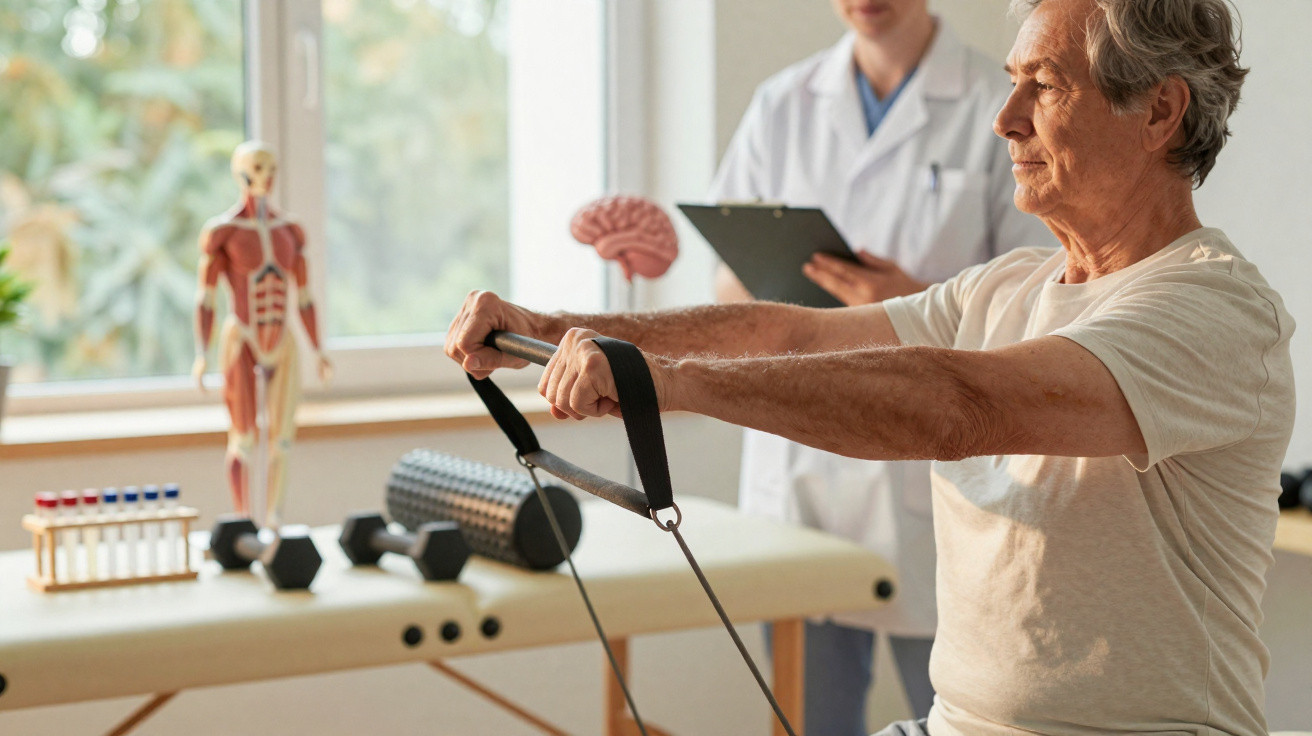
For decades, nearly every experimental medicine has targeted the brain directly. A newer strand of research now indicates that the muscles in our arms and legs may affect how long memory remains intact, even after the established signs of Alzheimer’s have appeared.
Muscles sending messages to the brain
Skeletal muscle was once regarded simply as the body’s mechanical engine: it contracts and enables movement, nothing more. That understanding is changing. Muscles also function as an endocrine organ, releasing signalling substances into the bloodstream whenever they contract.
Known as myokines, these signalling molecules travel well beyond the muscle where they originate. They can reach the liver, fatty tissue, the immune system – and the brain.
A key protein in this research is cathepsin B, whose levels commonly increase following physical activity. Earlier studies in animals and people associated higher cathepsin B levels with clearer thinking and stronger learning ability.
Exercise makes muscles speak a chemical language, and parts of that language seem to promote learning, memory and brain plasticity.
Plasticity refers to the brain’s capacity to adjust: strengthening or weakening connections between neurons and creating new ones. This mechanism supports the formation of new memories and the retention of skills over time.
Testing a radical idea in an Alzheimer’s model
Researchers chose to test the idea at its most extreme. Rather than directing treatments at amyloid plaques in the brain – the sticky deposits characteristic of Alzheimer’s – they attempted to amplify messages produced by muscle itself.
They worked with mice genetically modified to develop Alzheimer-like changes in the brain and memory difficulties as they grew older. Scientists used a viral vector to introduce genetic instructions into the muscles of some animals. Acting as a minute delivery vehicle, the vector instructed muscle cells to produce additional cathepsin B.
Importantly, the virus was engineered to act only in muscle tissue. The researchers did not manipulate the brain directly.
Six months later: the brain looks damaged, the memory less so
After six months, treated and untreated mice showed a marked difference. Mice whose muscles produced more cathepsin B performed substantially better in spatial-memory tests. In certain assessments, their learning performance came close to that of healthy, non-Alzheimer mice of the same age.
The team then examined the hippocampus, the seahorse-shaped brain area that is essential for making new memories. In untreated Alzheimer-model mice, the creation of new hippocampal neurons – called neurogenesis – normally falls sharply. In the treated animals, this reduction was mostly reversed.
Despite the brain still showing disease markers, the machinery for making new neurons and flexible synapses had switched back on.
Protein patterns also changed in the brain, muscles and blood. Protein-expression profiles in treated mice became more similar to those in healthy animals, pointing to a wider resetting of biological processes associated with memory and cellular repair.
A route that bypasses classic Alzheimer’s targets
One of the most notable results concerned what remained unchanged. Following months of treatment, the usual hallmarks of disease were still present. Amyloid deposits had not disappeared, and evidence of inflammation in the brain could still be detected.
Nevertheless, behaviour improved. This disconnect challenges the assumption that amyloid must be cleared in order to preserve memory.
Instead, cathepsin B seems to alter the brain’s response to damage. It increases proteins linked to synaptic plasticity, protein production and neurogenesis. Put simply, it appears to help the brain compensate for injury rather than remove it.
A double-edged molecule
The findings are not simple. Researchers obtained a different result when they increased cathepsin B in healthy mice without Alzheimer-like disease. Those animals developed memory difficulties.
The same molecule that supports a vulnerable brain may disrupt a healthy one when pushed too far.
This difference indicates that cathepsin B may be a helper whose effects depend on context, rather than a universally beneficial cognitive enhancer. It could be helpful only when brain circuits are already under strain and harmful when they are operating normally.
What this could mean for future Alzheimer’s treatments
This research contributes to a wider change in Alzheimer’s studies: considering the body as an interconnected system rather than viewing the brain in isolation. Messages from muscle, fat tissue, the gut and immune cells may all influence how resilient the ageing brain remains.
Focusing on muscle instead of neurons offers several attractive possibilities for drug development. Muscle tissue is more accessible, simpler to biopsy and less fragile than brain tissue. Treatments might be administered as muscle injections or as systemic therapies designed to selectively increase particular myokines.
Strategies currently being considered include:
- Medicines that safely raise beneficial myokines such as cathepsin B only when required
- Compounds that mimic exercise by activating muscle signalling without demanding intense workouts
- Gene therapies intended to adjust muscle-to-brain communication in people at high risk
- Personalised exercise plans informed by blood measurements of myokine levels
Researchers also emphasise that results in mice take time to translate into human treatments. Dosage, long-term safety and the possibility of cognitive side effects in healthy people all require close investigation.
Where exercise fits into the picture
These findings provide biological support for advice neurologists have repeated for years: remaining physically active generally benefits brain health. Regular activity encourages muscles to release a mixture of myokines, not only cathepsin B, which appear to support neurons and blood vessels.
Various forms of activity may produce different combinations of signals. Aerobic activities, including brisk walking, cycling and swimming, are often associated with better blood flow and elevated levels of certain growth factors. Strength training activates major muscle groups and may alter how muscles store and use energy.
| Type of activity | Typical effect on body | Relevance to brain research |
|---|---|---|
| Aerobic exercise | Raises heart rate and circulation | Linked with greater blood flow to the brain and myokines associated with neurogenesis |
| Strength training | Builds and maintains muscle mass | Helps sustain greater muscle “endocrine” output over time |
| Light daily movement | Cuts down prolonged sedentary time | Could deliver frequent, smaller pulses of muscle-derived signals |
For those concerned about dementia risk, researchers often advise combining cardiovascular exercise, strength training and balance exercises with good sleep habits, social interaction and mental challenges such as acquiring new skills or languages. These measures act through different biological mechanisms that together influence brain resilience.
Key concepts behind the muscle–brain link
Some terminology used in this research may seem abstract. Two concepts are particularly important to this muscle-focused approach to Alzheimer’s.
Myokines: Small proteins released by muscle cells during contraction. They can affect appetite, inflammation, metabolism and brain function. Cathepsin B is one of many myokines; others, including irisin, have also been linked with cognitive benefits in animals.
Neurogenesis: The production of new neurons from stem-like cells, occurring mainly in the adult hippocampus. Although it takes place on a modest scale compared with early life, these new cells appear to aid adaptable learning and mood regulation. Exercise, stress, diet and inflammation can each either promote or suppress the process.
A future clinic appointment offers one way to imagine how this could work in practice. Someone in their late fifties with a strong family history of Alzheimer’s might one day undergo not just a brain scan, but detailed blood testing for myokines and other peripheral markers. Rather than receiving one medicine directed at the brain, they could leave with a combined plan: an individualised exercise programme, potentially a muscle-focused therapy, and ongoing checks of changes in both muscle signals and memory-test results.
Risks must also be considered. Excessively stimulating pathways such as cathepsin B in people without neurodegeneration could damage memory or affect other organs. Unequal availability of gene therapies or costly biological medicines might deepen existing health inequalities. Nor would a muscle-based treatment remove the need to address established brain changes in later-stage disease.
Still, the central message is unexpectedly encouraging: memory may not be determined by the brain alone. Muscle strength and activity, along with the chemical messages muscles release, may form part of a broader approach to keeping Alzheimer’s at bay for longer.

















Comments
No comments yet. Be the first to comment!
Leave a Comment